| |
المؤلفون / Authors
الملخص / Abstract
الكلمات المفتاحية / Keywords
أقسام الملف
Introduction
Material and methods
Results and Discussion
Conclusions
References
|
| An Event Related Potential (ERP) for Detection of the (P300) Brain Waves in Elder People Based on IoMT |
| Auns Q. H. Al-Neami 1 , Qayssar A. Ahmed 2 Fatimah Haitham Abdulateef 3 |
| Rana Idan Abed (4) |
| 1,2,3University of Al-nahrain , Iraq-Baghdad, |
| 4 Ministry of Health, Iraq-Baghdad |
| |
| |
| Abstract |
|
| The current study intends to construct a system helps elder people who suffers from Alzheimer’s disease (AD) at present time. The system designed to detect those which may have mental issues that could developed into (AD) in the future, where the early detection of symptoms are greatly helps in the treatment procedure. Two devices built to fulfills this aim, the first one work as assisted device helping (AD) patients and their caregivers in daily activities throughout; (monitoring, tracking, remaindering), remotely by the use of internet of medical things (IoMT) technology, and showed results in displaying location with direction and speed in real time, also the reminder succeeded in timing up to 95%, the same success found in the detection of fall in different angles. The second device focuses on the early detection of Mild Cognitive Impairment (MCI) which could developed in the future into serious mental disorder such as (AD). The design based on the event related potential (ERP) principles and the latency of its (P300) wave component to detect the presence of latent peak in the neural response and use it as a non- invasive diagnosis tool. |
|
| Keywords: Alzheimer, P300, Diagnosis, Monitoring, Assistance, Cognitive, IoMT . |
|
| |
|
| |
|
| |
|
| |
|
| 1.Introduction |
|
| Alzheimer’s disease (AD) is defined as progressive, irreversible brain disorder that gradually destroys thinking skills as well as memory, by other words limits the ability to carry out the normal daily tasks [1]. The name of the disease derived from the name of Dr. Alois Alzheimer. Dr. Alzheimer found changes in the brain of a dead woman by unusual mental disease. The symptoms listed as memory loss, language problems, and unpredictable behavior [2, 3]. |
|
| The patho-physiology of (AD) is related to different factors such as: The extra-cellular deposition of beta-amyloid plaques, Accumulation of intracellular neurofibrillary tangles, Oxidative neuronal damage, inflammatory cascades [4]. |
|
| |
|
|

|
|
| Figure (1) The Effects of (Tangles) and (Plaques) in Brain [5]. |
|
| Mild cognitive impairment (MCI) presents a transitional state in cognitive functions between changes deriving from the aging process and those which classify as Dementia and Alzheimer’s disease [6]. |
|
| 2.Material and Methods |
|
| The biomedical system presented in the current study are contains two separated devices one for assisting and the other for diagnosis. |
|
 |
|
| Figure (2) the Biomedical System Devices. |
|
| 2.1.Biomedical Assistance Device |
|
|
The biomedical assistance device aims to help elder peoples with mild (AD) stages to depends on their own in completing daily activities, and on the other hand give their caregivers to monitor them remotely when they are far from them. The circuit design is shown in figure (3). As seen the device contains four units; sensing unit, control unit, display unit, and power unit. The sensing units contain two types of sensors, motion sensing unit which contain gyroscope and accelerometer, and a location sensor used for it a global positioning sensor.
|
|
 |
|
|
Figure (3) The Assistance Device Circuit Diagram.
|
|
| For control unit two microcontrollers are attached to the design one to provide data controlling while the other provide the wireless communication to the caregiver’s internet of things platform. The display unit is important because it display the date and time like normal watch in addition to displaying the reminders messages of important events sat by the caregiver. The power unit is a rechargeable battery to supply the circuit with necessary power. |
|
| The geographical location display property in the assistance device is depends on the (GPS) technique. The module used is (Neo- 6m Module) as shown in figure (4). This module is a (GPS) receiver with a built in ceramic antenna which provides a strong satellite search capability. It has signal indicator, that help to monitor the status of the module. The red indicator continuously flashes telling that the module is in the search stage, while when it receives a reliable signal it goes off. With its backup battery, the module can save the data when the main power is shut down accidentally. Personal computer (PC) is used to program the microcontroller connected to the GPS module which is NodeMCU ESP8266. After the connections were done, software uploaded to the microcontroller. |
|
 |
|
| Figure (4) The (GPS) Module. |
|
| The program loaded to the microcontroller is first ensures the Wi-Fi connection between the microcontroller and the IoMT platform through the specific Authentication number and Wi-Fi credentials (network name & password). After this the program checks the number of satellites signals available in the current locations in case of at least three signals received by the GPS module the software will store the information of: latitude, longitude, speed and direction in addition to the number of satellites discovered by the module [7]. Gyroscope provides information about the angular velocity of the three axes (X, Y, Z) and the accelerometer provides values of the angle of inclination in three axes, by combining these two modules together an efficient tracking tool can be achieved [8]. The benefit of this tool in the current study is detecting the condition of falling on ground that the patient may pass through. The motion processing unit (Gy521 & ADXL335) continuously calculate the data of angular velocity and acceleration in three dimensions, and finding (Ө and φ) angles of (X and Y) axes respectively in degrees through the code uploaded to the microcontroller. |
|
|
A threshold angles in (X and Y) axes are defined; if the angles threshold in one or both axes are crossed a fall case can be identified. In (X) axis the threshold angles are chosen in the range that greater than (25o) and less than (100o), while in (Y) axis the same range were used and additional threshold added that lie between (300o to 340o) to confirm more fall probability. The angles are continuously calculated and transmitted from the Arduino Nano to the Node MCU ESP8266 that supports the Wi-Fi connection with the IoMT platform. In cases of crossing the threshold a fall condition identified and an automatic notification appears in the caregiver’s smart phone. One of the major challenges that (AD) patients faces daily is the forgetfulness. The reminder is one feature of the Assisted device, this reminder displays real date and time and reminding massages for medication times. For a reminder design LCD (2X16) display and Buzzer connected with the microcontroller. The display is connected by (I2C) protocol which is serial protocol to connect devices using only two weirs SCL (serial clock) and SDA (serial data). The software uploaded will be responsible for displaying the current time and date of the current location and this information will be taken from the internet as an internet of thing platform used it’s easier and more accurate to be synchronized and up to date anytime, anywhere to the time zone the (AD) patient uses the assistance. Buzzer was connected to act as alarming sound to draw attention of patient to the time of taking pills. The connection between the assistance device and the caregiver is through the use of internet of things technology. Many free mobile applications support this technology, the one used in this study was the (Blynk) application. This application makes a link between it and between the project for sending and receiving information. This link through an authentication credentials (user name, password, and authentication number).

|
|
| 2.2.Biomedical Diagnosis Device |
|
| The diagnosis device work principles are shown in figure (5) while its circuit diagram is shown in figure (6). |
|
 |
|
| Figure (5) Diagnosis Device Work Principles. |
|
 |
|
| Figure (6) Diagnosis Device Circuit Diagram. |
|
| The diagnosis device of (MCI) can assist physicians in detection of the (MCI) by the recording of event related potential with an audio stimulation for the brain waves and analyze it, to use the results of analysis to locate the (P300) component latency. the physician can diagnose if the patient has MCI which may progress to (AD) or not. The diagnosis circuit designed by using surface electrodes, instrumentation amplifiers (AD623 & TLC2252), microcontroller, in addition to speakers for the audio stimulation. For software design a Spike recorder software used as signal recorder, the two circuits (main and stimulation circuit powered by 5V). Three electrodes are used, two for the EEG signals and one electrode used as a reference electrode, see Figure (7). |
|
 |
|
| Figure (7) electrode fixing cap’s two surfaces (a) outer, (b) inner. |
|
| Two microcontrollers are used in the design, one microcontroller is attached to the circuit manage the output of the signals that acquired, amplified and filtered, in order to be processed by it. The second microcontroller is responsible for the generating the standard and oddball tons along the test period. These tones work as an audio stimulation. The response of the audio stimulation by the brain is translated as a P300 wave generated by the parietal lobes of the brain. A series of amplification processes are used to the brain signals that obtained by electrodes. |
|
| First one is the amplifying with gain of four times (4x Gain): amplifier is used to amplify very low signals, eliminating noise and interference. This is done by the use of the (AD 623) amplifier. This amplifier has the feature of flexibility in setting the gain with one resister. It’s gain easily programed from (1-1000), (G = 1 with no external resister). The second phase of amplification is by the use of Band-pass filter (BPF) with gain amplifier, the (TLC2252) operational-amplifier used. |
|
| The third phase is an another gain amplifier using the potentiometer that can sets the gain from (1-10x). The potential reference of the amplifiers useful in defining the zero output voltage. Its usefulness appears in cases of bipolar signals amplification, because it used for providing a ground voltage virtually. The voltage on the reference terminal can be varied from −Vcc to +Vcc in the current study it is chosen equal Vcc/2. |
|
| 2.2.1Electrode Setup and Testing EEG waves |
|
| Oliver et al. [27] |
|
| 1.The electrodes were placed over (P3-P4) locations using usual EEG (10-20) system, as shown in figure (8 left). |
|
| 2.Gel added below the electrodes at the top of the head cap. ensuring there is little hair contacting electrodes with skin. |
|
| 3.Ground electrode placed over the mastoid process. Figure (8) right. |
|
 |
|
| Figure (8) Electrode positions (left) P3 and P4 Electrodes (right) Ground. |
|
| 4.The electrodes connected by connectors to the circuit, as illustrated in figure (9). |
|
 |
|
| Figure (9) Connecting the Electrodes with the Circuit. |
|
| 5.Now running the recording software on the computer, setting the filters, and reduce electrical noise as possible. |
|
|
6.Make the subject being tested to hold and relax in attempting to record a clear EEGs without noises.

|
|
| 3.Results and Discussion |
|
| 3.1.Results of Assistance Device |
|
| The outdoor locating or (GPS) module is tested to locate the position of the Assistance device and as a results the location of the (AD) patient who wear the device. The results show the location of the device on a map with displaying the important information like the speed, direction, number of satellites participating in locating and of course the latitude and longitude. Figure (10) shows the screen of the (IoMT) platform or the Blynk application used to display the locating property. |
|
 |
|
| Figure (10) IoMT platform shows the location of the assistive device. |
|
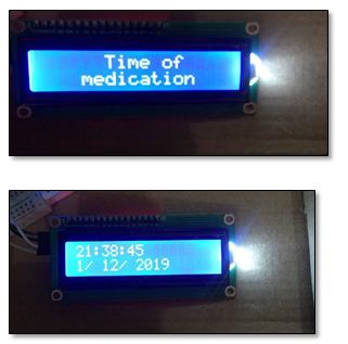
| Timing property of the device is for displaying the current date and time. The time is taken from the internet to be accurate with the time zone, and work like watch. Figure (11 (top)) shows the LCD displaying the time and date. A time of medications is also displayed on the LCD display as reminder in specific times set by the caregiver using the Blynk application figure (11)(button)) |
|
 |
|
| Figure (11) LCD displays date (button) and time (top) |
|
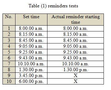
| A test of (10) reminders are test and the results are listed in the table (1): |
|
 |
|
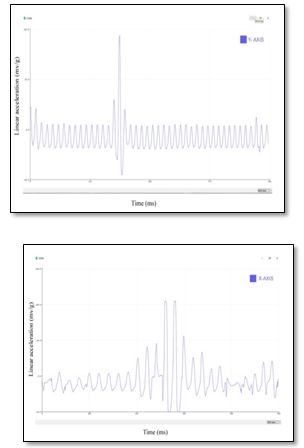
| The motion processing unit represented by the gyroscope and accelerometer follows the status of the device. For sense the tilt or achieve the fall detection property, besides the gyroscope the accelerometer attached in the design. As in the gyroscope the accelerometer tested first and displays its acceleration curve of the three directions (X, Y, Z) on the serial plotter. Figure (12) shows the acceleration curve and its change in acceleration due to tilt. The (left) it shows the linear acceleration change of X-axis due to tilt with angle (60o). While in (right) the acceleration change at tilt with angle (80o) in Y-axis is showed. |
|
 |
|
| Figure (12) linear acceleration change of X-axis due to tilt (top)with angle (60o), (button) with angle (80o). |
|
|
The fall detection was informed to the caregiver through the Blynk notification, alarming him that the patient had a problem and needs help.

|
|
| 3.2.Results of Diagnosis device: |
|
| 3.2.1.Results of the Audio Stimulation: |
|
| Audio stimulation used to active a stimulation in the subject’s brain to generate a response that translated as P300 wave. This audio stimulation wave was shown in figure (13). Figure shows the screen of the software displaying the standard tone that streaming through the test (yellow wave) and the oddball tone which randomly appears through the test (blue wave). |
|
 |
|
| Figure (13) Spike recorder setting window. |
|
| 3.2.2.Results of P300 Wave |
|
| In order to check the location or the latency of P300 wave a record of EEG is done with an audio stimulation. A group of (8) male subjects with no hearing problems, with ages range from (31-62) years old apply the test in a room with less electrical noise as possible. The subjects relaxed on a chair with hand rest and asked to count 50 oddball stimulations. First of all, a record of EEG is tested to judge the reality of the wave and be sure it’s an EEG wave no other types of wave signals or noise, to do that a subject asked to close his eyes for a while (10 seconds or less) and notice the wave’s amplitude were it must be enlarges with eye enclosure, comparing it with the eyes open. This test is shown in figure (14). |
|
 |
|
| Figure (14) ensuring the EEG wave legality. |
|
| After checking the wave signal is real EEG signal, and audio stimulation then started to generate the audio stimulation and from the software two new channels were chosen to display the standard and oddball tones. At this point the display must show channels and waves as in figure (15). |
|
 |
|
| Figure (15) the three channels displaying (EEG, standard, and oddball tones). |
|
| As previously mentioned the test is done on (8) males subjects with good hearing sense and after recording their event related potentials with Spike recorder software and analyze it to find P300 curves and check its latency, the results were four figures first was the neural response to the standard tone, second was the neural response to the oddball tone, while the third one was Monte Carlo curve, the final one was the combining of the three figures (Standard, oddball, Monte Carlo curves) were as shown in figure (16): |
|
 |
|
| Figure (16) (EEG, standard tone, oddball tone) of subject (45) years old. |
|
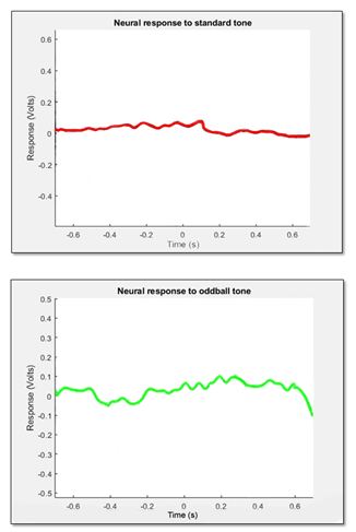
| The neural response of standard tones and the neural response of oddball tones are demonstrated in figure (17). |
|
| |
|
 |
|
| Figure (17) Neural response of standard tones (top), Neural response of oddball tones (button). |
|
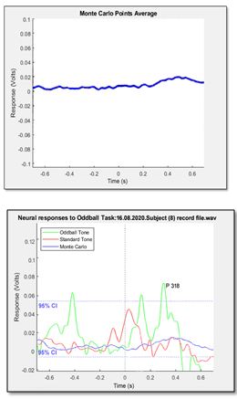
| The Monte Carlo curve and the neural response curves with P300 peak are shown in figure (18). |
|
 |
|
|
Figure (18) Monte Carlo curve (top), Neural response curves with P300 peak (button).

|
|
| 4.Conclusions |
|
| This study intends to design and implement a system for helping people in both early detection of Mild cognitive impairment (MCI) or the early onset of Alzheimer’s disease (AD), and assisting people suffering from dementia specially (AD) with its early stages to face daily challenges as independents as possible. The early detection device follow the concepts of the event related potential (ERP) with audio stimulation, locating the P300 component waveform and use it as a noninvasive diagnosing tool for the early detection of (MCI). In the assistance device the locating feature through (GPS), works better in open fields than in closed areas. The direction and speed were well displayed on the (IoMT) platform and its updating were satisfying to produce a simple locating map function. A reminder was tested and gave a results accuracy up to 95% and it’s found to be highly related to strength of internet connection in area were the assistance used. The motion tracking feature mainly used to reflect the stability when the subject works, and avoiding the cases of fall without knowing by the caregiver. The feature fond to cover the angles of abnormal position of an elder people. The call button found to be an easy, efficient, fast and simple way to call for a help or need for something from the caregiver remotely. Finding the (P300) or the highest peak of the oddball for diagnosis present a clear way to notice it’s latency with time. The user will only look to the curve and find the highest peak and find its latency on the x-axis, then comparing the value of (P300) with the normal range and use it in the diagnosis. With this system, both the assistance and diagnosis devices are providing a help for an important segment of the society that serve the country for long years, with low cost, reliable and easy to use devices that makes the user more confident and less dependent and live life with less obstacles of aging. |
|
| 5.References |
|
| 1." Basics of Alzheimer’s Disease and Dementia" “what is Alzheimer Disease” - National institute aging. web. May 16, 2017 <https://www.nia.nih.gov/health/what-alzheimers-disease> |
|
| 2.Al-Shaban S., Al-Neami A., Al-Shaban M., “Non- Linear Principal Component Analysis Neural Network for Blind Source Separation of EEG Signals”, Research Journal of Applied Sciences, Engineering and Technology 2(2): 180-190, ISSN: 2040-7467(2010). |
|
| 3.Demetrius L., Magistretti P., Pellerin L., “Alzheimer’s disease: the amyloid hypothesis and the Inverse Warburg effect”, Hypothesis and theory article published:14 January (2015) doi: 10.3389/fphys.2014.00522. |
|
| 4.Wujek J. R, Dority D., Frederickson R.C.A. And Brunden K. R., “Deposits of AβFibrils are Not Toxic to Cortical and Hippocampal Neurons in Vitro”, Neurobiology of Aging, Neurobiology of Aging, Vol. 17, No. 1, 0197-4580/96 pp. 107-113, (1996). |
|
| 5.Ashfaq M., Talreja N., Chuahan D., Srituravanich W. “Carbon Nanostructure-Based Materials: A Novel Tool for Detection of Alzheimer’s Disease”. In: Ashraf G., Alexiou A. (eds) Biological, Diagnostic and Therapeutic Advances in Alzheimer's Disease. Springer, Singapore. https://doi.org/10.1007/978-981-13-9636-6_4 (2019). |
|
| 6.Marina M. S., “P300 Evoked Potential in patients with Mild cognitive impairment”, ACTA Inform Med., 21(2):89-92., (2013). |
|
| 7.Kandel E., Schwartz J. “Principles of Neural Science, 4th ed.”, United State of America: McGraw-Hill., (2000), p.324. |
|
| 8.Saba M., Auns Q. H., “Controlled Wheelchair System Based on Gyroscope Sensor for Disabled Patients”, Biosciences Biotechnology Research Asia, Vol. 15(4), P. 921-927, December (2018). |
|
 |
|
| |
|
| |
|
| |
|
| |
|
| |
|
| |
|